F003
Side of Tumor Center
Anatomical laterality of the tumor center relative to the brain midline: left, right, or midline.
Advancing structured neuro-oncology reporting through standardized annotation, machine-readable findings, and clear dataset documentation.
Un progetto per la refertazione strutturata in neuro-oncologia tramite annotazione standardizzata, reperti leggibili automaticamente e documentazione chiara del dataset.
The resulting paper will be submitted to Nature. Contributing annotators whose reports pass the quality check will be included as co-authors.
Il paper risultante verrà sottomesso a Nature. Gli annotatori i cui referti superano il controllo qualità saranno inclusi come co-autori.
| Label | Description |
|---|---|
| ET | Enhancing Tumor |
| NETC | Non-Enhancing Tumor Core |
| ED | Edema |
| TC | Tumor Core, defined as ET + NETC |
| WT | Whole Tumor, defined as ET + NETC + ED |
| ID | Name | Description | Assigned To |
|---|---|---|---|
| F001 | Tumor Center Region | The tumor center is defined as the barycenter, or center of mass, of the TC region. The location of this point within the atlas is used to assign the corresponding anatomical region. To avoid cases in which the barycenter lies on a boundary, a spherical neighborhood with a radius equal to one tenth of the axial diameter is considered. The assigned region is then defined as the most prevalent region within this sphere. | Machine |
| F002 | Tumor Center Coordinates | The coordinates of the barycenter are computed as the mean coordinates of all voxels in the TC region. | Machine |
| F003 | Side of Tumor Center | Defined as the anatomical laterality of the tumor center based on the position of the TC barycenter relative to the brain midline, e.g., left, right, or midline. | MachineAnnotator |
| F004 | Eloquent Brain Involvement | Determined using the atlas, this field specifies which of the following eloquent functional areas are involved by the tumor: vision, motor, speech motor, and speech receptive. | Machine |
| F005 | Enhancement Quality | Qualitative assessment of CET contrast enhancement, indicating whether enhancement is present and how pronounced it appears. | Annotator |
| F006 | Proportion of ET | Automatically derived from the segmentation mask using the formula: n. voxelET / n. voxelWT. | Machine |
| F007 | Proportion of ED | Automatically derived from the segmentation mask using the formula: n. voxelED / n. voxelWT. | Machine |
| F008 | Proportion of NETC | Automatically derived from the segmentation mask using the formula: n. voxelNETC / n. voxelWT. | Machine |
| F009 | Thickness of ET Margin | Defined as the geometrically computed average thickness of the ET margin. | Machine |
| F010 | Definition of the Enhancing Margin | Assesses whether most of the outer margin of the enhancement is well defined or poorly defined. | Annotator |
| F011 | Definition of the Non-Enhancing Margin | Assesses whether most of the outer margin of the non-enhancing component is well defined or poorly defined. | Annotator |
| F012 | Intratumoral Hemorrhage | Assesses whether intrinsic hemorrhage is visible anywhere in the tumor matrix. | Annotator |
| F013 | TC Size | Defined as the largest perpendicular cross-sectional diameters of the TC across the axial, sagittal, and coronal planes. | Machine |
| F014 | ED Size | Defined as the largest perpendicular cross-sectional diameters of the ED across the axial, sagittal, and coronal planes. | Machine |
| F015 | Tumor Heterogeneity | The degree of heterogeneity of the internal architecture of the tumor. | Annotator |
| F016 | TC Affected Areas | Determined using the atlas, this field specifies which brain regions are affected by the TC. | Machine |
| F017 | ED Affected Areas | Determined using the atlas, this field specifies which brain regions are affected by the ED. | Machine |
| F018 | Number of Lesions | Number of independent lesions observed in the brain. | MachineAnnotator |
| F019 | Type of Lesion | Categorical variable indicating the observed lesion type. Possible values: single, multicentric, or multifocal. | MachineAnnotator |
| F020 | Ependymal Invasion | Assesses whether the TC abuts any adjacent ependymal surface. | Annotator |
| F021 | Depth of TC | Determines the depth of the TC. Possible values: cortical, subcortical white matter, deep white matter and basal nuclei. | MachineAnnotator |
| F022 | Tumor Center Macroarea | The macroarea, or lobe, in which the barycenter lies, obtained using the atlas. | MachineAnnotator |
| F023 | Midline Shift | The shift of the midline caused by the tumor mass, in millimeters. | Annotator |
| F024 | Tumor Shape | The approximate shape of the tumor mass. | Annotator |
| F025 | Tumor Origin | Where the tumor originates, following the grades: Periventricular, Gyral, or Deep white matter. | Annotator |
| F026 | Tumor Crosses Midline | State whether the tumor crosses the midline. | Annotator |
| F027 | Subject specific findings | Any subject specific findings that do not fall under any predefined category. | Annotator |
Free-text reporting instructions and annotator-facing fields translated from the Italian annotation guide.
Reports should be compiled in free-text format, evaluating each relevant template field. Information that is not present should be omitted, avoiding explicit statements about the absence of findings or characteristics that are not described.
During annotation, it is recommended to follow the same evaluation progression across cases to ensure consistency. Measurements should always be reported in millimeters. In cases with multiple lesions, the annotation protocol should be repeated for each lesion so that the reported information clearly refers to the correct lesion.
| ID | Field | Clinical Meaning |
|---|---|---|
| F003 | Side of Tumor Center | Anatomical laterality of the tumor center relative to the brain midline: left, right, or midline. |
| F005 | Enhancement Quality | Qualitative assessment of CET contrast enhancement, including whether it is present and how pronounced it appears. |
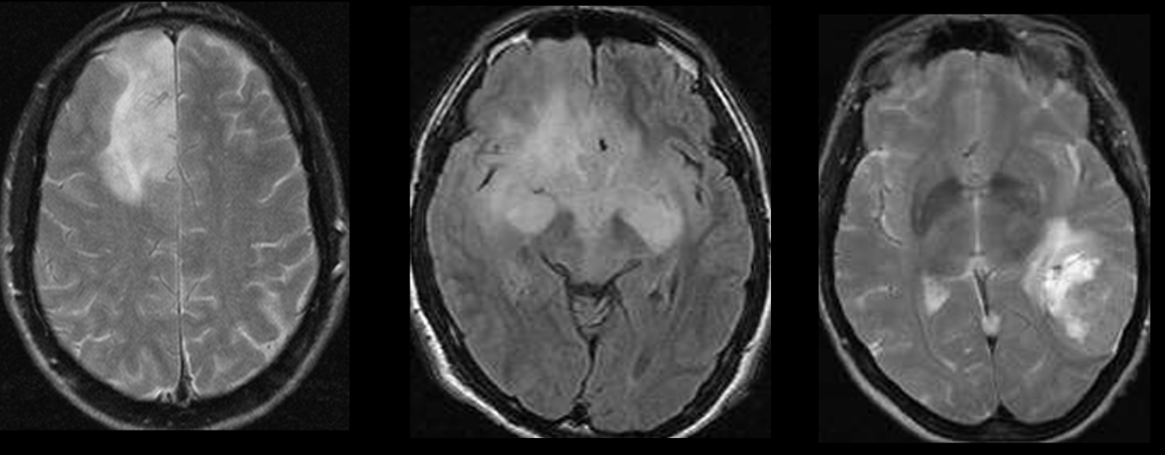
| F010 | Definition of the Enhancing Margin | Whether most of the outer enhancing margin is well defined or poorly defined. |
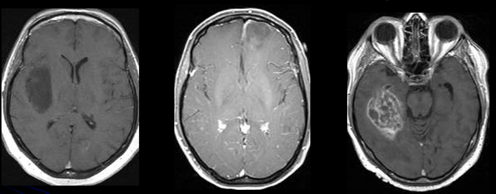
| F011 | Definition of the Non-Enhancing Margin | Whether most of the outer non-enhancing component margin is well defined or poorly defined. |
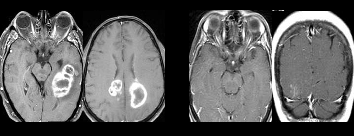
| F012 | Intratumoral Hemorrhage | Whether intrinsic hemorrhage is visible anywhere in the tumor matrix. |
| F015 | Tumor Heterogeneity | Degree of heterogeneity of the tumor internal architecture. |
| F018 | Number of Lesions | Number of independent lesions observed in the brain. |
| F019 | Type of Lesion | Categorical lesion type: single, multicentric, or multifocal. |
| F020 | Ependymal Invasion | Whether the TC abuts an adjacent ependymal surface. |
| F021 | Depth of TC | Possible values: cortical, subcortical white matter, deep white matter, and basal nuclei. |
| F022 | Tumor Center Macroarea | The macroarea or lobe in which the barycenter lies. |
| F023 | Midline Shift | Shift of the midline caused by the tumor mass, reported in millimeters. |
| F024 | Tumor Shape | Approximate shape of the tumor mass. |
| F025 | Tumor Origin | Likely origin category: periventricular, gyral, or deep white matter. |
| F026 | Tumor Crosses Midline | Whether the tumor crosses the midline. |
| F027 | Subject-Specific Findings | Any subject-specific finding that does not fall under a predefined category. |
Anatomical laterality of the tumor center relative to the brain midline: left, right, or midline.
Qualitative assessment of CET contrast enhancement, including whether it is present and how pronounced it appears.
Whether most of the outer enhancing margin is well defined or poorly defined.
Whether most of the outer non-enhancing component margin is well defined or poorly defined.
Whether intrinsic hemorrhage is visible anywhere in the tumor matrix.
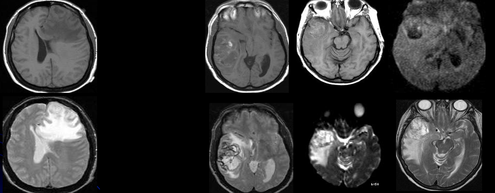
Degree of heterogeneity of the tumor internal architecture.
Number of independent lesions observed in the brain.
Categorical lesion type: single, multicentric, or multifocal.
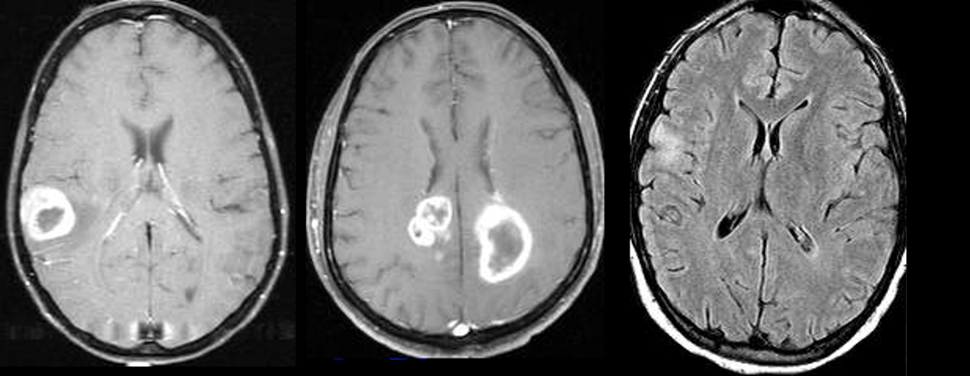
Whether the TC abuts an adjacent ependymal surface.
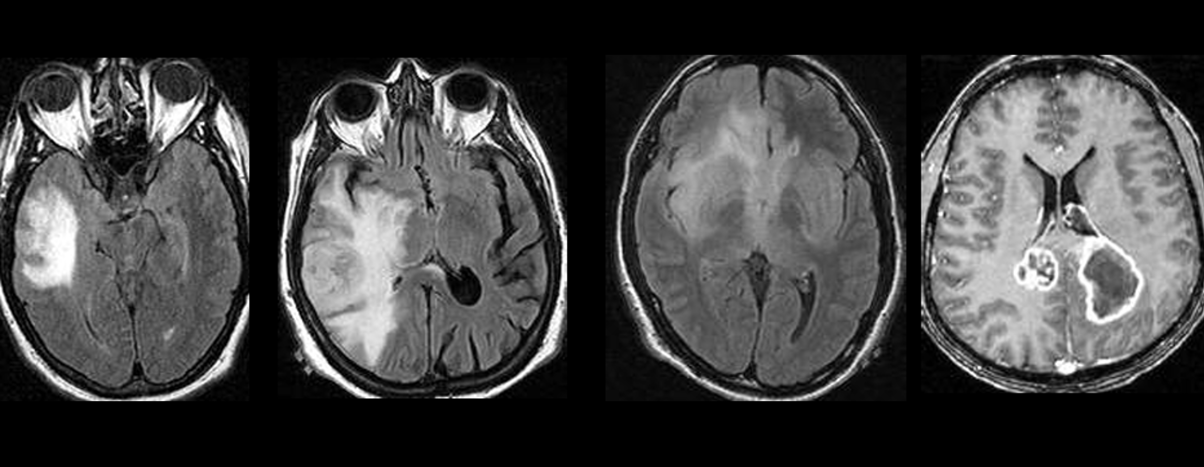
Possible values: cortical, subcortical white matter, deep white matter, and basal nuclei.
The macroarea or lobe in which the barycenter lies.
Shift of the midline caused by the tumor mass, reported in millimeters.
Approximate shape of the tumor mass.
Likely origin category: periventricular, gyral, or deep white matter.
Whether the tumor crosses the midline.
Any subject-specific finding that does not fall under a predefined category.
Definizione strutturata dei campi del dataset per la refertazione su ToothFairy4M.
| Etichetta | Descrizione |
|---|---|
| ET | Enhancing Tumor |
| NETC | Non-Enhancing Tumor Core |
| ED | Edema |
| TC | Tumor Core, definito come ET + NETC |
| WT | Whole Tumor, definito come ET + NETC + ED |
Definito come la lateralità anatomica del centro del tumore in base alla posizione del baricentro del TC rispetto alla linea mediana del cervello, ad esempio sinistra, destra o linea mediana.
Valutazione qualitativa dell'enhancement del contrasto CET, indicando se l'enhancement è presente e quanto appare pronunciato.
Valuta se la maggior parte del margine esterno dell'enhancement è ben definita o scarsamente definita.
Valuta se la maggior parte del margine esterno della componente non-enhancing è ben definita o scarsamente definita.
Valuta se un'emorragia intrinseca è visibile in qualsiasi punto della matrice tumorale.
Il grado di eterogeneità dell'architettura interna del tumore.
Numero di lesioni indipendenti osservate nel cervello.
Variabile categorica che indica il tipo di lesione osservata. Valori possibili: singola, multicentrica o multifocale.
Valuta se il TC è a contatto con una superficie ependimale adiacente.
Determina la profondità del TC. Valori possibili: corticale, sostanza bianca sottocorticale, sostanza bianca profonda e nuclei della base.
La macroarea, o lobo, in cui ricade il baricentro, ottenuta utilizzando l'atlante.
Lo spostamento della linea mediana causato dalla massa tumorale, in millimetri.
La forma approssimativa della massa tumorale.
Dove origina il tumore, secondo le categorie: Periventricolare, Girale, Sostanza bianca profonda.
Indica se il tumore attraversa la linea mediana.
Qualsiasi reperto specifico del soggetto che non rientra in alcuna categoria predefinita.
I referti vanno compilati in formato testuale libero, valutando ciascun campo del template. Le informazioni non presenti devono essere omesse, evitando di riportare esplicitamente l'assenza di reperti o caratteristiche non descritte.
Durante l'annotazione è consigliabile seguire sempre la stessa progressione di valutazione, così da garantire coerenza tra i diversi casi. Le misure vanno sempre riportate in millimetri.
In caso di lesioni multiple, il protocollo di annotazione deve essere ripetuto per ciascuna lesione, in modo da rendere sempre chiaro a quale lesione si riferiscono le informazioni riportate.
Nel protocollo sono definiti criteri specifici e oggettivi per classificare una lesione come singola, multifocale o multicentrica. ED è indicato in rosso, NETC in verde ed ET in blu.
Definiamo come multicentrica una lesione in cui i corrispettivi ED sono separati, mentre multifocale nel caso non lo fossero.
Il referto di esempio rappresenta il modello da seguire per la compilazione.
In sede parasagittale destra, in corrispondenza della regione cingolata posteriore/precuneale a prevalente interessamento cortico-sottocorticale, si documenta una singola formazione espansiva intrassiale a morfologia ovalare, a segnale disomogeneamente ipointenso in T1 e iperintenso in T2.
La lesione presenta componente centrale necrotica e orletto solido periferico con enhancement disomogeneo post-contrastografico, in parte lineare e in parte nodulare, intenso. Si associa enhancement piale.
È riferito marginale interessamento della porzione posteriore del corpo calloso in sede destra da parte del core tumorale. Le alterazioni iperintense in T2/FLAIR perilesionali, estese alla sostanza bianca profonda dei lobi parietale e occipitale destri, si propagano inoltre nel corpo calloso, tratto posteriore e medio in sede destra, con minimo sconfinamento controlaterale a sinistra a livello del corpo calloso posteriore.
La massa determina effetto espansivo con obliterazione degli spazi liquorali periencefalici in sede occipito-parietale posteriore destra e compressione del trigono, del corno occipitale e della porzione posteriore del corpo del ventricolo laterale destro. Si associa deviazione della linea mediana, setto pellucido, di circa 2 mm verso sinistra. Il ventricolo laterale sinistro presenta morfovolumetria conservata; III e IV ventricolo regolari. Non segni di erniazione.
Coesiste importante quadro di sofferenza microangiopatica cronica biemisferica.
Questa scomposizione mostra come ciascun campo del template trovi una corrispondenza nel testo libero.
| ID | Campo | Finding |
|---|---|---|
| F003 | Lato del Centro del Tumore | "In sede parasagittale destra", "regione cingolata posteriore/precuneale" e "parietale e occipitale destri". |
| F005 | Qualità dell'Enhancement | "enhancement disomogeneo post-contrastografico, in parte lineare e in parte nodulare, intenso". |
| F010 | Definizione del Margine Enhancing | "orletto solido periferico con enhancement disomogeneo post-contrastografico". |
| F011 | Definizione del Margine Non-Enhancing | Assente nel referto, in quanto non ben definito. |
| F012 | Emorragia Intratumorale | Assente nel referto. |
| F015 | Eterogeneità del Tumore | "segnale disomogeneamente ipointenso in T1", "componente centrale necrotica" e "orletto solido periferico". |
| F018 | Numero di Lesioni | Una sola lesione: "si documenta una singola formazione espansiva". |
| F019 | Tipo di Lesione | Singola: "si documenta una singola formazione espansiva". |
| F020 | Invasione Ependimale | Assente nel referto. |
| F021 | Profondità del TC | "a prevalente interessamento cortico-sottocorticale". |
| F022 | Macroarea del Centro del Tumore | "regione cingolata posteriore/precuneale" e "sede occipito-parietale posteriore destra". |
| F023 | Spostamento della Linea Mediana | "deviazione della linea mediana, setto pellucido, di circa 2 mm verso sinistra". |
| F024 | Forma del Tumore | "formazione espansiva intrassiale a morfologia ovalare". |
| F025 | Origine del Tumore | Probabile origine girale/cortico-sottocorticale. Evidenza testuale: "regione cingolata posteriore/precuneale a prevalente interessamento cortico-sottocorticale". |
| F026 | Il Tumore Attraversa la Linea Mediana | "minimo sconfinamento controlaterale a sinistra a livello del corpo calloso posteriore". |
| F027 | Reperti Specifici del Soggetto | "Si associa enhancement piale", "marginale interessamento della porzione posteriore del corpo calloso", "effetto espansivo con obliterazione degli spazi liquorali periencefalici" e "importante quadro di sofferenza microangiopatica cronica biemisferica". |
La piattaforma di annotazione è disponibile su yggdrasil.ing.unimore.it.
Tutto il lavoro di refertazione verrà fatto sulla piattaforma Yggdrasil.
Per qualsiasi problema, contattare reportx@unimore.it.
Ecco un breve tutorial: